Jumat, 23 Maret 2012

Somatic stem cells obtained from skin cells for first time ever

March 22, 2012 08:05 PM

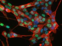
This is an immunofluorescence microscopy image of the induced neural stem cells (iNSCs) using antibodies against two neural stem cell markers SSEA1 (red color) and Olig2 (green color).
Breaking new ground, scientists at the Max Planck Institute for Molecular Biomedicine in Münster, Germany, have succeeded in obtaining somatic stem cells from fully differentiated somatic cells. Stem cell researcher Hans Schöler and his team took skin cells from mice and, using a unique combination of growth factors while ensuring appropriate culturing conditions, have managed to induce the cells' differentiation into neuronal somatic stem cells. "Our research shows that reprogramming somatic cells does not require passing through a pluripotent stage," explains Schöler. "Thanks to this new approach, tissue regeneration is becoming a more streamlined - and safer - process.
Up until now, pluripotent stem cells were considered the 'be-all and end-all' of stem cell science. Historically, researchers have obtained these 'jack-of-all-trades' cells from fully differentiated somatic cells. Given the proper environmental cues, pluripotent stem cells are capable of differentiating into every type of cell in the body, but their pluripotency also holds certain disadvantages, which preclude their widespread application in medicine. According to Schöler, "pluripotent stem cells exhibit such a high degree of plasticity that under the wrong circumstances they may form tumours instead of regenerating a tissue or an organ." Schöler's somatic stem cells offer a way out of this dilemma: they are 'only' multipotent, which means that they cannot give rise to all cell types but merely to a select subset of them - in this case, a type of cell found in neural tissue - a property, which affords them an edge in terms of their therapeutic potential.
To allow them to interconvert somatic cells into somatic stem cells, the Max Planck researchers cleverly combined a number of different growth factors, proteins that guide cellular growth. "One factor in particular, called Brn4, which had never been used before in this type of research, turned out to be a genuine 'captain' who very quickly and efficiently took command of his ship - the skin cell - guiding it in the right direction so that it could be converted into a neuronal somatic stem cell," explains Schöler. This interconversion turns out to be even more effective if the cells, stimulated by growth factors and exposed to just the right environmental conditions, divide more frequently. "Gradually, the cells lose their molecular memory that they were once skin cells," explains Schöler. It seems that even after only a few cycles of cell division the newly produced neuronal somatic stem cells are practically indistinguishable from stem cells normally found in the tissue.
Schöler's findings suggest that these cells hold great long-term medical potential: "The fact that these cells are multipotent dramatically reduces the risk of neoplasm formation, which means that in the not-too-distant future they could be used to regenerate tissues damaged or destroyed by disease or old age; until we get to that point, substantial research efforts will have to be made." So far, insights are based on experiments using murine skin cells; the next steps now are to perform the same experiments using actual human cells. In addition, it is imperative that the stem cells' long-term behaviour is thoroughly characterized to determine whether they retain their stability over long periods of time.
"Our discoveries are a testament to the unparalleled degree of rigor of research conducted here at the Münster Institute," says Schöler. "We should realize that this is our chance to be instrumental in helping shape the future of medicine." At this point, the project is still in its initial, basic science stage although "through systematic, continued development in close collaboration with the pharmaceutical industry, the transition from the basic to the applied sciences could be hugely successful, for this as well as for other, related, future projects," emphasizes Schöler. This, then, is the reason why a suitable infrastructure framework must be created now rather than later. "The blueprints for this framework are all prepped and ready to go - all we need now are for the right political measures to be ratified to pave the way towards medical applicability."
Source : Max-Planck-Gesellschaft

http://www.biologynews.net/archives/2012/03/22/somatic_stem_cells_obtained_from_skin_cells_for_first_time_ever.html

Study and Removal of the Frog's Brain

Name:__________________________________________
brain


Starting at the most anterior part of the head, the olfactory nerves connect to the nostrils and then to the olfactory lobes (A) where odors are processed. Just posterior to the olfactory lobes are two elongate bodies with rounded bases, this is the cerebrum (B), and it is the frog's thinking center. The cerebrum is the part of the brain that helps the frog respond to its environment. Posterior to the cerebrum are the optic lobes (C), which function in vision. The ridge just behind the optic lobes is the cerebellum (D), it is used to coordinate the frog's muscles and maintain balance. Posterior to the cerebellum is the medulla oblongata (E) which connects the brain to the spinal cord (F).
Brain Part
Function
Letter
Cerebellum

 
Cerebrum

 
Olfactory Lobe

 
Optic Lobe

 
Medulla Oblongata


Removal of the Frog's Brain

Turn the frog dorsal side up. Cut away the skin and flesh on the head from the nose to the base of the skull. With a scalpel, scrape the top of the skull until the bone is thin and flexible. Be sure to scrape AWAY from you. With your scalpel held almost horizontally, carefully chip away the roof of the skull to expose the brain. Use scissors to cut away the heavier bone along the sides of the brain.
To receive extra credit for removing the brain, you must present it to me on a paper towel. All lobes should be attached with as much tissue as possible present. GOOD LUCK!

FROG BONES

The bones of the frog follow the same pattern (basically) as other vertebrates. The lower leg of the frog is a muscular leg that the frog uses for jumping. There are 3 main sets of bones in the lower leg. The femur is found in the upper thigh, and the tibiofibula is found in the lower part of the leg. The foot and ankle are made of the tarsals and metatarsals (toes).
To expose the frog's leg bones you must remove the thigh muscle - the biceps femorus and the calf muscle - the gastrocnemius. You can leave the Achilles tendon intact (this tendon connects the muscle to the bone). The tarsals and metatarsals do not need to be exposed.
To receive extra credit, clean your bones by removing the surrounding muscle. Bring the two bones on a paper towel. Good Luck!
Label the leg bones: Femur, Tibiofibula, tarsals, metatarsals, pelvic girdle
Other Frog Dissection Pages


http://www.biologycorner.com/worksheets/frog_brain_leg.html

Sabtu, 17 Maret 2012

Bakteri Berkomunikasi Lewat Sentuhan

Suka dengan artikel ini?

Jelajahi artikel-artikel FaktaIlmiah yang berdasarkan apa yang dibaca dan ditonton teman-teman.
Terbitkan aktivitas Anda sendiri dan dapatkan kendali penuh.
Login


Minggu, 4 Maret 2012 - Bagaimana jika bakteri dapat bicara satu sama lain? Bagaimana jika mereka punya indera sentuhan? Sebuah studi terbaru dari para peneliti UC Santa Barbara menunjukkan keduanya benar, dan berteori kalau sel demikian, pada faktanya, perlu berkomunikasi untuk melakukan fungsi tertentu.

Penemuan ini muncul baru saja dalam jurnal  Genes & Development.
Christopher Hayes, asisten professor biologi molekuler, seluler, dan perkembangan UCSB, bekerjasama dengan mahasiswa pasca sarjana  Elie Diner, Christina Beck, dan Julia Webb untuk mempelajari  uropathogenic E. coli (UPEC), yang menyebabkan infeksi saluran kemih pada manusia. Mereka menemukan hubungan mirip saudara antara system sel yang telah lama diduga sebagai musuhnya.
 Makalah mereka menunjukkan kalau bakteri menunjukkan sebuah system hambat pertumbuhan tergantung kontak (Contact–Dependent Inhibition – CDI) yang dapat menghambat bakteri tanpa system demikian hanya bila bakteri targetnya memiliki CysK, sebuah enzim metabolic yang dibutuhkan untuk sintesis asam amino cysteine. CysK ditunjukkan berikatan dengan racun CDI — sebuah enzim yang memecah  RNA ó dan mengaktifkannya.
Untuk sebuah system sel yang diduga hanya ada untuk membunuh bakteri lain – seperti diduga pada CDI – hasil ini mengejutkan, kata Hayes, karena ini berarti sel penghambat CDI+ harus mendapatkan izin dari targetnya sebelum melakukan tugasnya.
 “Ini pada dasarnya bermakna sel penghambat bertanya pada sel target, “bolehkah saya menghambat anda?” jelas beliau. “Ini tidak masuk akal. Mengapa menambah lapisan kompleksitas baru? Mengapa menambah factor izin? Ini temuan yang tidak biasa.
 “Kami pikir sekarang system CDI tidak dibuat semata karena sel-sel ini ingin keluar dan membunuh sel lain,” lanjut Hayes. “Hasil kami menunjukkan kemungkinan kalau sel-sel ini menggunakan CDI untuk berkomunikasi sebagai saudara dan bekerjasama; misalnya, dalam membentuk biofilm, yang memberikan kekuatan dan kelangsungan hidup yang lebih baik pada bakteri.”
 Studi ini menunjukkan enzim CysK sebagai katalis potensial untuk komunikasi bakteri tersebut – seperti jabat tangan rahasia, atau sebuah password. Secara sederhana, kata Hayes, “Jika anda memiliki sandi yang tepat, anda diizinkan bergabung; sebaliknya, anda akan diusir.”
Walaupun hanya UPEC yang dipelajari dalam makalah ini, Hayes mengatakan kalau temuan ini berpotensi untuk berlaku pada pathogen lainnya mulai dari meningitis bakteri hingga wabah, serta bakteri berbasis tanaman yang dapat merusak pertanian.
David Low, Profesor biologi molekuler, seluler, dan perkembangan serta pengarang kedua dalam makalah ini, menjelaskan karya laboratorium Hayes sebagai terobosan penting dalam bagaimana bakteri berkomunikasi – dan penerapan praktisnya yang suatu saat dapat diwujudkan.
 “Kita baru saja mulai mendapatkan petunjuk kalau bakteri mungkin bicara satu sama lain dengan bahasa yang tergantung kontak,” kata Low. Mereka bersentuhan dan merespon satu sama lain dalam cara berbeda tergantung system CDI dan factor genotype lainnya. Harapan kami adalah pada puncaknya karya ini dapat membantu pengembangan obat yang memblokir atau meningkatkan komunikasi tergantung sentuhan, untuk bakteri berbahaya ataupun yang menguntungkan.”
 Penelitian ini didukung oleh dana dari  National Institutes of Health dan National Science Foundation.
Sumber berita:
Referensi jurnal:
E. J. Diner, C. M. Beck, J. S. Webb, D. A. Low, C. S. Hayes. Identification of a target cell permissive factor required for contact-dependent growth inhibition (CDI). Genes & Development, 2012; DOI: 10.1101/gad.182345.111

http://www.faktailmiah.com/2012/03/04/bakteri-berkomunikasi-lewat-sentuhan.html

Sabtu, 10 Maret 2012

Kebersamaan Ketika Raker JSC 2012

    Sabtu kemarin pada tanggal 10 Maret 2012 telah dilaksanakan raker JSC 2012. Raker diadakan di Gazebo Perpus Pusat pukul 07.00 WIB. Suasana  raker yang santai menjadi suatu kebersamaan yang menyenangkan.
    Raker JSC telah dihadiri oleh Ketua HIMA BIOLOGI periode 2012-2013, Mahbub Masduqi. Selain Ketua HIMABIO, hadir pula perwakilan dari CBF (Cempaka Bio Farm), Ketua GC (Green Community), dan BSC.
    Acara inti Raker yaitu pemaparan progja masing-masing bagian yang dimulai dari PH (Pengurus Harian), Biro PSDM, Departemen Keilmiahan, Departemen, Informasi dan Humas, Departemen JEC (Jasmina English Club). Bukan sekedar lembaran kertas, progja yang telah dibacakan dan dikritisi hingga akhirnya memperoleh kesepakatan akan menjadi langkah awal dari masing-masing bagian untuk menjalankan masing-masing progjanya selama satu periode kepengurusan tahun 2012.
    Diakhir Raker, JSC juga membahas tentang Mipashow yang rencana akan diadakan bulan Maret ini. Tentunya acara ini akan sangat menarik dan sudah ditentukan temanya hlo, PENASARAN??? tunggu info selanjutnya.
   Akhir kata sebagai penyemangat untuk kawan-kawan JSC'ers, tetap SEMANGAT untuk melaksanakan Progja-Prohja yang telah disusun dan lakukan sebaik mungkin.

Kamis, 01 Maret 2012

Raker JSC 2012

JSC Be The Winner !

Salam Hangat dan semangat.
      Sebentar lagi JSC akan melaksanakan Raker periode 2012-2013 yang tentunya akan membahas program kerja masing-masing bagian baik PH, Biro PSDM, dan semua departmen.
      Nah, program kerja atau yang biasa disebut progja ini merupakan rancangan kegiatan/aktivitas yang akan dilakukan selama satu periode kedepan sekaligus memberi tanggung jawab kepada pengurus masing-masing bagian.
      When and Where???
Tidak lama lagi raker JSC dilaksanakan, yaitu tanggal 10 Maret 2012 bertempat di Markas JSC tercinta alias Gazebo Perpus Pusat, hehehe.
So, jangan sampai ketinggalan acaranya. See you next at D-Day.